


Please enter the answer below before you can view the full text.
7+6=
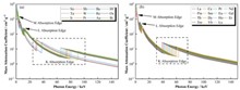
BackgroundWith the extensive application of nuclear technology in medical field, the demand for radiation protection materials has been increasing. Traditional lead-based protective materials suffer from disadvantages such as heavy weight and biological toxicity, making the development of lightweight, lead-free and flexible X-ray protective materials a research priority.PurposeThis study aims to investigate the X-ray shielding mechanism and develop high-performance flexible protective materials suitable for medical diagnostic X-ray tubes operating within voltages below 150 kV.MethodsFirstly, the mass attenuation coefficients of different elements were calculated using XCOM program, revealing complementary K-edge absorption effects among tungsten (W), bismuth (Bi), and gadolinium (Gd) elements across different energy ranges. Secondly, Monte Carlo simulations were employed to predict the shielding performance of W, Bi, and Gd element combinations for X-ray tubes operating under 120 kV tube voltage with 2.7 mm Al filtration. Finally, flexible protective materials comprising W+Bi-based composite layered with Gd-based materials were prepared and experimentally tested.ResultsExperimental test results show that the optimized composite material achieves excellent shielding performance with shielding efficiency of 82.98%, mass attenuation coefficient of 5.86 cm2·g-1, linear attenuation coefficient of 9.16 cm-1 and half value layer of 0.08 cm. The effective atomic number (Zeff) analysis confirms enhanced absorption in the 50~90 keV range, which corresponds to the typical energy range of medical diagnostic X-rays.ConclusionsResults of this study demonstrate that the material design strategy based on complementary K-edge absorption effects can effectively improve the performance of flexible X-ray protective materials to shield both bremsstrahlung and characteristic radiation in the X-ray spectrum, providing valuable insights for developing next-generation lightweight, lead-free, and flexible X-ray protective materials for medical applications.
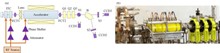
BackgroundElectron beam injectors are the core components that determine the performance of free electron laser terahertz sources (FEL-THz). Radio frequency (RF) accelerator-based injectors can generate high-quality electron beams required to drive high-power THz radiation.PurposeThis study aims to design and commission a compact Free Electron Laser for Terahertz (FEL-THz) injector using an Independently Tunable Cell (ITC) structure, which balances high quality, cost-effectiveness, and compactness.MethodsThe collaborative FEL-THz system developed by Huazhong University of Science and Technology and the University of Science and Technology of China, was taken as research object. Initially, optimization concepts and design results based on the ITC structure were introduced. Thereafter, the system stabilization, beam diagnostics and system optimization measures were detailed for beam commissioning and operation. Relevant schemes and research validation were completed through beam dynamics simulations and online experiments.ResultsThe experimental results demonstrate that the optimized ITC injector achieves an electron beam energy of approximately 13.7 MeV with an energy spread of 0.41% and a transverse emittance of 8.8 mm mrad, meeting high-power and miniaturization requirements.ConclusionThe simulation and experimental results both indicate that, through optimization design combined with indirect diagnostic techniques, the ITC injector can meet the demands for both high power and miniaturization of FEL-THz. This configuration holds promising potential for advancing the miniaturization of high-power FEL-THz facilities. Furthermore, the application of indirect diagnostic techniques can also compensate for the insufficient diagnostic methods during the commissioning process, laying the groundwork for further promotion of the ITC injector.
BackgroundVanadium oxide (VOx) detectors play a crucial role in the fields of space infrared detection and imaging due to their sensitivity to infrared radiation, particularly in the Long-Wave Infrared (LWIR) range, as well as their advantages of high sensitivity, low noise, uncooled operation, and cost-effectiveness. However, performance degradation caused by space ionizing radiation, particularly the decrease in responsivity and the increase in noise levels, poses a significant threat to the imaging quality and reliability of these detectors.PurposeThis study aims to bridge the current gap in research on the space radiation effects on VOx detectors and provide input for their radiation hardening.MethodsGround-based radiation simulation experiments were conducted to analyze the radiation damage to VOx detectors. Both online and offline testing methods were employed to systematically investigate the changes in detector output under varying radiation doses. Additionally, post-irradiation annealing tests were performed to examine the specific patterns of performance degradation in VOx detectors under space radiation environments.ResultsThe results indicate that space radiation significantly affects the output performance of VOx detectors. When the accumulated dose reaches 31 krad(Si), the non-uniformity of the detector's blackbody temperature response and the number of dead pixels surge. At 39 krad(Si), the non-uniformity of the blackbody response escalates to 88%, the dead pixel rate climbs to 66%, and the output image becomes aberrant. Within 24 h after the irradiation test, the annealing effect of the VOx infrared array detector is evident, with the average grayscale value of the blackbody response output closely aligning with the pre-irradiation results after 72 h of annealing, and the number of dead pixels tending towards 0 after one week of annealing.ConclusionsSpace radiation significantly impacts the output of VOx detectors, with the detector's output performance declining as the radiation dose accumulates, reaching a point where the device cannot recognize objects in images at 24 krad(Si). The total ionizing dose effect at lower accumulated dose values does not constitute permanent damage, as the detector's performance can be restored through room-temperature annealing. This study provides valuable insights for the subsequent design of radiation-hardened VOx detectors for space applications.
BackgroundThe β-ray surface density measurement instrument is widely used for the online measurement of the surface density of electrode sheets in the coating process of lithium batteries. A significant measurement error was observed when the source-to-film distance (SFD) varied under fixed source-to-detector distance (SDD).PurposeThis study aims to investigate and minimize the impact of SFD variations on the accuracy of β-ray surface density measurements.MethodsFirstly, experimental tests were conducted using the β-ray surface density measurement device with a fixed SDD to quantify the influence of SFD changes (4~8 mm) on copper film surface density measurements. Then, Geant4 Monte Carlo simulations were performed to model β-ray scattering effects under three collimator configurations: detector-side single collimator, source-side single collimator, and dual collimators. Finally, the optimized source-side single collimator geometry was experimentally validated by reproducing SFD variations and recalculating measurement deviations.ResultsInitial experimental results reveal that 1 mm SFD variation causes a maximum measurement deviation of 13.3% in surface density values. Monte Carlo simulation results demonstrate that the source-side single collimator configuration minimizes scattering effects, showing the smallest mean deviation among all three geometries tested. Experimental validation confirms this improvement with the maximum deviation decreasing from 13.3% to 3.5% after device optimization.ConclusionsResults of this study show a 73.7% reduction in measurement error caused by SFD variations is achieved by adopting a source-side single collimator, providing a practical solution for enhancing online surface density measurement accuracy in lithium battery production.
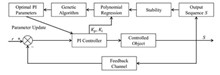
BackgroundThe Hefei Advanced Light Facility (HALF) is a fourth-generation synchrotron radiation source based on a diffraction-limited storage ring. Its electron beam energy is 2.2 GeV, with an emittance target of less than 100 pm·rad which requires stability within 5×10⁻⁵ for the magnet steady-state power supply and reduces overshoot in step response when changing the current operating point. Due to the need for over a thousand magnet steady-state power supplies at HALF, the empirical tuning method using conventional proportional-integral-derivative (PID) controller parameters requires a significant amount of time.PurposeThis study aims to design a PID controller parameter auto-tuning algorithm for the magnet steady-state power supply of HALF, to achieve optimal PI parameters.MethodsBased on polynomial regression and genetic algorithms, an auto-tuning algorithm for PI controller parameters was developed for the magnet steady-state power supply controller. Then, BUCK circuit structure was adopted for the magnetic steady current power supply, and the simulation and physical experiment verification of the prototype of the magnetic steady current power supply were carried out. Finally, this algorithm was integrated with the supervisory computer system to record various data points. Development and testing of this algorithm were conducted on the magnet steady-state power supply to verify the stability of the output current.ResultsThe test results of the power supply step response and output current stability show that the step response achieves a smooth transition, and the rising speed is increased several times. The output current stability is within 1×10⁻⁵. The key technical indicators meet the operation requirements and significantly improve the debugging efficiency.ConclusionsThe algorithm proposed in this paper provides an effective method for improving the debugging efficiency of large-scale power supplies in the future.
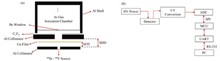
BackgroundActive neutron interrogation (ANI) measurement systems can quantify the fissile masses of special nuclear materials (SNMs) using neutrons and are widely used in nuclear safeguarding fields. However, different hydrogen densities effect the delayed neutron count rate differently, and the hydrogen-containing matrix in the waste drum weakens the signal of delayed neutrons and limits the fissile mass measurement precision of the ANI system.PurposeThis study aims to improve the assay performance of the ANI system in nuclear safeguard measurements by developing a matrix correction method.MethodsBased on the response of the flux monitor, a new matrix correction method was developed based on the conventional correction methods. A WM3210 PAN Shuffler system model was constructed using the Geant4 toolkit, and correction method was explored for common matrix materials. Finally, U3O8 materials with different enrichments and distributions were applied to the effectiveness comparison between new correction method and the traditional method.ResultsThe results show that for U3O8 materials with different enrichments and distribution states, both the traditional correction method and the new correction method can effectively reduce the influence of the matrix material on the measurement of SNM mass. For the case where U3O8 material is located at the center of the matrix, the average relative deviation of the 235U mass obtained by the new correction method is 13.6%, while that of the traditional method is 23.8%. For the case where U3O8 material is uniformly dispersed in the matrix, the average relative deviation of 235U mass obtained by the new correction method is 7.78% whilst that of the traditional method is 20.0%.ConclusionResults of this indicate that the new method demonstrates better correction capability than the traditional one.
BackgroundMost of the irradiators that undertake the fundamental dosimetry studies are installed with radioactive sources of high-specific activity, which are only approved to be transported by qualified containers before the onsite loading being performed.PurposeThis study aims to design a novel irradiator featuring triple screen functionality to comply with the international and domestic regulation on the transportation of the radioactive source.MethodsFirstly, based on the precise construction of the irradiator, a comprehensive simulation was conducted to analyze the control characteristics of the irradiation device on the beam according to the accurate modelling for the irradiator. Then, detailed simulation and analysis was performed in order to obtain the properties of the field in terms of radiation dosimetry research interests. Finally, a novel irradiator featuring triple screen functionality was implemented to facilitate the onsite installation and the following work.ResultsThis novel irradiator is appropriate to perform the onsite loading for the radioactive source. The scattered components in the spectrum can be reduced by 25% with the presence of a scattering chamber in the irradiator. The leakage radiation in the normal working area can be decreased by 7 orders of magnitude due to the triple screening functionality of the irradiator, the optimization is proposed for installation of the key components in the irradiator. The deformation of the spectra in the radiation field caused by the collimator complex is about 0.2%. The wall effect in the course of the absolute measurement of air kerma for three types of typical ionization chamber is calculated in the 60Co γ radiation field, and the correction factors diverge less than 0.2% from the ones in the earlier publications.ConclusionsThe irradiator designed and implemented in this study can be a proper candidate to support the radiation dosimetry related work under the current regulation. The compact structure enables the irradiator to provide an extended dynamic range of dose rate, leading to a relatively long period of service.
BackgroundThe Shanghai High-repetition-rate XFEL and Extreme Light Facility (SHINE) is characterized by high brightness, full coherence, and high repetition rates. For focusing high-repetition-rate hard X-rays, compound refractive lenses (CRL) must withstand significant heat loads, which can potentially lead to optical performance failure due to high thermal stress.PurposeThis study aims to optimize the maximum allowable repetition rate of X-ray beams while maintaining thermal stress within permissible limits to ensure reliable optical performance of CRLs under high-repetition-rate operation.MethodsFirstly, a finite element method (FEM) was employed to perform thermal-structural coupled analysis of beryllium CRLs with various internal spherical radii (0.3 mm, 1.0 mm, 2.0 mm, and 5.8 mm) under different photon energies (5 keV, 7 keV, and 15 keV). Then, the maximum thermal stress and temperature distributions were systematically evaluated under the initial 1 MHz repetition rate condition. Finally, the repetition rates were optimized to keep the maximum thermal stress below the permissible threshold of 120 MPa (50% of beryllium's yield strength), with special focus on the central region of the lens where stress concentration occurs.ResultsAnalysis results show that CRLs with internal radius R=0.3 mm experience a maximum thermal stress of 1 008 MPa at 7 keV photon energy at 1 MHz repetition rate, far exceeding the permissible limit. After optimization, the maximum thermal stress is reduced by 93% to 74 MPa by lowering the repetition rate to 100 kHz. The maximum temperature decreases from 201 °C to 36.6 °C, an 82% reduction. For larger radius CRLs (R=5.8 mm), the optimized repetition rate could be increased to 500 kHz for 7 keV X-rays while maintaining thermal stress below the threshold.ConclusionsBy optimizing the repetition rate according to CRL geometry and beam energy, thermal stress can be effectively controlled within safe limits. Increasing the internal spherical radius of CRLs from 0.3 mm to 5.8 mm allows for higher repetition rates (from 100 kHz to 500 kHz for 7 keV photons), thereby enhancing operational capabilities while ensuring reliable optical performance and structural integrity.
BackgroundThe length of the photon beam transportation of Shanghai High repetitioN rate XFEL and Extreme light facility (SHINE) exceeds 1 km, the angular vibration of reflecting mirrors would be magnified at the endstations due to the long-distance transportation.PurposeThis study aims to explore the possibility of suppressing the angular vibration by using a dynamic vibration absorber.MethodsAccording to the big pitch angular vibration of the mirror system at 14.6 Hz, a dynamic vibration absorber was designed with variable stiffness aiming at suppressing the vibration of mirror system for SHINE.ResultsBy tuning the nature frequency of the dynamic vibration absorber designed in this study at 14.6 Hz, results show that the vibration of reflecting mirrors at 14.6 Hz is reduced from 154 nard to 27.9 nrad, while vibrations at other frequencies are not disturbed, the total vibration is improved from 193.2 nrad to 76.9 nrad.ConclusionsThis indicated that the certain angular vibration of the mirror system could be suppressed by dynamic vibration absorber, which provided a new solution for angular vibration control of the advanced X ray light sources.
BackgroundThe energy selection system (ESS) is a key part of the cyclotron-based proton therapy facility. In addition to regulating the energy of the fixed-energy proton beam generated by the cyclotron to match the depth of the tumor, the other main role is to control the beam emittance to meet the therapeutic requirements. Existing schemes for controlling the emittance are based on switching combinations of collimators with different apertures, where the adjustable emittance is limited by the number of combinations of apertures, and the need for a small aperture collimator upstream or downstream of the ESS to suppress the beam intensity limits the possibility of increasing the high-energy beam intensity.PurposeThis study aims to propose the beam optics design of proton therapy energy selection system (ESS) on the basis of the superconducting cyclotron CYCIAE-230 under development by the China Institute of Atomic Energy (CIAE) that can flexibly control the beam emittance and transmission.MethodsFirst of all, the main optical design features of this ESS was described, and the Monte Carlo method based FLUKA code was applied to calculating the beam emittance, energy, and energy spread of a proton passing through a degrader with a set of collimators. Then, beam optical software TRANSPORT and TURTLE were combined to design the linear beam optics with consideration of placing a set of width-adjustable slits in front of the achromatic section and combining the beam optical conditions at the corresponding positions to control the beam divergence. Finally, the feasibility of flexible and adjustable beam emittance and transmission efficiency for esigned ESS was verified by calculations.Results & ConclusionsThe results show that the ESS meets the demand of proton therapy, and the rational use of adjustable slit can make the ESS have the characteristics of flexible and adjustable beam emittance, so as to realize the adjustable beam transmission in the energy selection section. This simple and flexible optical design proposed in this study for adjusting the state of the beam can be used for regulating the beam intensity for proton therapy and other research applications, which has a wide range of application prospects.
BackgroundLarge-volume container waste generated from nuclear facility decommissioning exhibits extremely inhomogeneous nuclide distribution. Its activity measured using the traditional tomographic gamma scanning (TGS) technique shows errors of up to six orders of magnitude; this hinders its safe disposal.PurposeThis study aims to develop an improved tomographic gamma scanning (ITGS) technique based on the equivalent surface source method and establish a reconstruction algorithm.MethodsFirstly, based on the count rate ratios of different γ-scan positions, the basic distribution of nuclides in the waste container was calculated, and the accurate activity reconstruction results were obtained by the combination of efficiency calibration. Then, γ-scan experiment was simulated for measuring various radioactive source distributions of Co-60 nuclides in large waste containers to verify the accuracy of this method, and the influence law for the nuclide distribution and measurement parameters on the reconstruction results was discovered.ResultsSimulation results demonstrate that a deeper radioactive source location or larger medium density can increase the activity reconstruction error. However, based on the same measurement time, the ITGS measurement error can be controlled within an order of magnitude compared with the conventional TGS.ConclusionThe proposed method achieves higher reconstruction accuracy under complex conditions such as multiple nuclide distributions and high medium density. Furthermore, it mitigates waste disposal safety hazards to humans and the environment by facilitating rational arrangements of the measurement system and activity reconstruction of radioactive waste containers.
BackgroundA fuel rod is a fundamental unit of a fuel assembly, and it directly impacts the safe operation of nuclear reactors. To efficiently detect internal defects in fuel rods, a high-resolution visual nondestructive testing method, X-ray imaging, i.e., digital radiography (DR), is employed.PurposeThis study aims to address the issue of low contrast in fuel rod X-ray DR images by proposing a brightness fusion and multiscale optimized enhancement algorithm.MethodsFirst, logarithmic and gamma transformations and further refined by incorporating local information fusion were employed to correct the brightness of fuel rod DR image. Subsequently, a wavelet function was applied for multiscale decomposition, enhancing and sharpening low-frequency components with Retinex, and non-local means (NL Means) was applied to filtering high-frequency components. Then, image enhancement was realized via wavelet reconstruction. Finally, quantitative analysis experiments were conducted using the DR images of fuel rods to evaluate the performance of the algorithm by means of two representative image quality assessment metrics, i.e., average gradient (AG) and information entropy (IE), and compared with that of new low-light image enhancement (NLIE) algorithm, homomorphic filtering (HMF) algorithm, and low light image enhancement (LIME) algorithm.ResultsThe experimental results demonstrate that quality of fuel rod DR image is significantly improved by image brightness fusion and multiscale optimized enhancement algorithm proposed in this study with the highest information entropy (IE) of 6.834 5, which is 10.2%, 3.3%, and 12.6% higher than NLIE, HMF and LIME algorithms, respectively, hence the internal defects of fuel rods are better highlighted.ConclusionsThis algorithm proposed in this study not only effectively improves the overall and local contrast of the fuel rod DR image, but also significantly highlights edge details, verifying its effectiveness in improving the quality of X-ray DR images.
BackgroundFacilitating object imaging through the utilization of cosmic-ray muons mandates the precise delineation of muon trajectories, where the pinpoint localization of muon impact points assumes paramount importance for effective muon track reconstruction. Existing muon track detection systems necessitate the integration of multifaceted electronic channels to attain meticulous positioning of muon impact points. The construction of such detection systems is distinguished by its intricacy and entails substantial associated costs.PurposeThis study aims to achieve a design for a muon track detection system that is characterized by simplicity, low cost, and high precision.MethodsThe Geant4 software was applied to the simulation of detectors comprising square and circular plastic scintillators coupled with silicon photon multipliers (SiPMs) without segmentation. The SiPMs was used to collect the number of photons and the time triggering SiPM responsed as characteristic parameters in the simulation, and a uncut square and circular plastic scintillator detector with an area of 200 mm × 200 mm was constructed, with a thickness of 10 mm. The surface was coated with a TiO2 reflective coating with a thickness of 0.11 mm and a reflectivity of 95%. Then, three types of artificial intelligence regression algorithms, i.e., extreme gradient boosting (XGBoost), multilayer perceptron (MLP) and long short-term memory (LSTM), were employed as the method for muon localization.ResultsThe simulation results demonstrate that LSTM algorithm achieves the highest accuracy among the three regression algorithms when photon number is considered as the characteristic parameter. Specifically, under the LSTM algorithm, the position resolution of a configuration comprising 12 SiPMs coupled to the upper surface of the detector can attain a resolution at the centimeter level. Furthermore, by employing photon number and trigger time as characteristic parameters, the position resolution of a setup involving only 6 SiPMs coupled to the side of the detector also reaches the centimeter level. Remarkably, these results align with the experimental findings obtained from a detector equipped with a photomultiplier tube (PMT) coupled to a large-area plastic scintillator.ConclusionsThis study employs the LSTM regression algorithm as the muon localization method, proposing a detector system structure for plastic scintillators with 6 SiPMs coupled to the side. The proposed structure is characterized by simplicity, low manufacturing cost, and achieves a positioning accuracy at the centimeter level.
BackgroundThe preparation of alumina ceramics is usually sintered under atmospheric pressure, and the sintering temperature is usually above 1 800 ℃ to achieve densification of alumina. High temperature and long-term heat preservation often make the grains of alumina ceramics overgrow, and it is impossible to predict what defects and porosity that occur inside the crystal, resulting in the decline of the comprehensive properties of alumina ceramics. Previous work predominantly focuses on studying the stable nano α-phase alumina (α-Al2O3) sintering process, microstructure, and abnormal grain growth, but few studies on the variations in microstructure and defects during the sintering process of metastable γ-phase alumina (γ-Al2O3), let alone use CaO as an additive in nanoceramic alumina.PurposeThis study aims to apply positron annihilation lifetime spectroscopy combined with X-ray diffraction (XRD) to investigating the impact of calcium oxide (CaO) addition on the sintering process of Al2O3 nanoceramics.MethodsFirstly, γ-phase nanoscale alumina powder with an average grain size of 20 nm, 99.99% pure and nano-calcium oxide powder with an average grain size of 160 nm, 98% pure was used as raw ceramic materials. Tablet samples of Al2O3 nanoceramics and Al2O3 nanoceramics doped with CaO (with a mass fraction of 1% CaO) were pressured under consistent experimental conditions. Then, the samples were sintered from 500 ℃ to 1 100 ℃ for 2 h, and cooling to room temperature. XRD was used to characterize the phases and compounds of the sample, and field emission scanning electron microscope (SEM) was employed to observe the microscopic morphology of the sample cross-section, accompanied by an EDS spectrometer for elemental composition analysis. Finally, a radioactive isotope 22Na with intensity of about 0.5×10⁶ Bq was used as positron source and fast-fast coincidence positron lifetime spectroscopy was applied to characterizing the sample defects. The total count of each lifetime spectrum was over 106, and all samples were measured for 2~3 times. The lifetime spectrum data were fitted and analyzed by LT(V9) program. The average value was taken as the result after spectral deconvolution.ResultsThe results of XRD and SEM show that the sintering process of CaO/Al2O3 nanoceramics is divided into two stages, no phase transition occurred from room temperature to 900 ℃, while significant phase transition occurred from 900 ℃ to 1 100 ℃. The addition of a small amount of CaO (such as 1% mass fraction) is uniformly distributed in the Al2O3 matrix at first, and with the increase of sintering temperature, it reacts with the Al2O3 to form a second phase and transform into a liquid phase at higher temperature. Analysis results of positron annihilation lifetime spectrum show that the size and number of vacancy clusters and micropores in CaO/Al2O3 nanoceramics before and after the phase transition are different from those of Al2O3 nanoceramics. In CaO/Al2O3 nanoceramics, vacancy clusters and micropores are more likely to form as the temperature rises, some micropores gradually merge to form macroscopic pores before phase transition whilst micropores in Al2O3 nanoceramics disappear with phase transition and grain growth, leaving only a small amount of micropores on the sample surface.ConclusionsThe addition of a small amount of CaO suppresses the growth of Al2O3 grains during the sintering process, resulting in a more uniform and denser structure. Simultaneously, the addition of CaO within Al2O3 nanoceramics induces the formation of a liquid phase. This alters the mass transfer mechanism from solid-state diffusion to liquid-phase flow, thereby delaying the phase transformation process of Al2O3 nanoceramics during sintering and leading to the formation of macroscopic pores.
BackgroundBipolar magnet power supplies using switching techniques are commonly used to provide stable excitation current for correction magnet coils. Specification of the Shenzhen Superconductive Soft-X-ray Free Electron Laser (S3FEL) project requires enhanced magnetic field stability of correction magnets. However, when switching power supplies are used for correction magnets at low currents, the very narrow pulse width of the power switch leads to poor stability in the output current.PurposeThis study aims to propose a method involving a series controllable bidirectional impedance circuit to improve the stability of low current output from the correction magnet power supply.MethodsFirstly, the characteristics of a controllable bidirectional impedance was identified when connected in series from the power supply to the correction magnet, and a Bode diagram was applied to analyzing its features. Then, a controllable bidirectional impedance circuit was designed on the basis of simulation analysis. Finally, experimental validation was conducted using the correction magnet and power supply from Shanghai Synchrotron Radiation Facility (SSRF).ResultsThe experimental results demonstrate that serially connecting controllable bidirectional impedances not only improves low current stability but also allows for smooth switching between MOSFETs and controllable bidirectional impedances.ConclusionsThe circuit design proposed in this study is simple and proves to be effective to improve the stability of bipolar magnet power supply under low current condition.
BackgroundBoron neutron capture therapy (BNCT) is a new type of binary targeted radiotherapy that is gradually entering the commercial stage. The accelerator-based boron neutron capture therapy (AB-BNCT) neutron sources used in hospitals are inevitably accompanied by neutron leakage and gamma-ray contamination during equipment operation and patient treatment.PurposeThis study aims to analyze the radiation safety of public during the operation of BNCT using Monte Carlo geometric splitting variance reduction technique.MethodsFirstly, based on a 10 mA 2.8 MeV proton accelerator neutron source, the International Commission on Radiological Protection (ICRP) and the basic standard for ionizing radiation protection and radiation source safety in China, GB 18871?2002 was taken as the reference standard for the annual effective dose limit of 1 mSv for the public. This dose limit was conservatively estimated to be equivalent to a dose rate limit of 0.5 μSv·h-1. Then, a water model with the size of 100 cm×30 cm×80 cm was placed at the outlet of beam shaping assembly (BSA) after neutron beam moderation and collimation, and the shielding calculations of AB-BNCT treatment room were conducted by Monte Carlo programs with various techniques. Finally, these results were comparatively analyzed to give shielding scheme design of the AB-BNCT treatment room, and the optimization design was carried out according to the weak link of the shielding.ResultsThe simulation results indicate that the variance reduction techniques of geometric splitting and Russian roulette for shielding simulation calculations has higher efficiency and achieve more accurate calculation results. After optimizing the shielding design of the BNCT treatment room, the conservative calculation results prove that the total dose rate at the corridor outside the treatment room is less than 0.5 μSv·h-1 when the shielding body is 60 cm thick boron containing heavy concrete, which meets the design requirements. In addition, the thickness range of the boron containing heavy concrete shielding wall required for radioactive workers is given to be 45~50 cm when the dose rate limit is less than 2.5 μSv·h-1.ConclusionsThe shielding design of BNCT treatment room using Monte Carlo geometric splitting variance reduction technique in this study is safe and reliable, providing a theoretical basis for the commercial development of BNCT.
BackgroundThere are a large number of Cloisonné enamel in the Palace Museum, among which there are many problems in the screening of the manufacture process and background information of the enamel with "Jingtai". X-ray technology can be applied to non-destructive test of the structure and composition of enamel, and help researchers to acquire the information behind cultural relics.PurposeThis study aims to apply X-ray techniques to the investigation of Cloisonné enamel in the Palace Museum for analyzing their period, glaze and structure.MethodsFirstly, two pieces of enamel inscribed with "Jingtai" in the Palace Museum were selected as the research object. Then, the X-ray Computed Tomography (CT), X-ray Fluorescence (XRF) and Raman spectroscopy techniques were applied to investigating the period, glaze, structure and composition of these two Cloisonné enamels. Finally, comparative analysis of these identified characteristics was conducted to infer the differences in different parts of Cloisonné enamels.ResultsAnalysis results indicate that both of two Cloisonné enamels are old utensils made by changing their shapes with recombination of the old pieces, thus forming a new piece of enamel with "Jingtai" engraved on the bottom.ConclusionsThrough the mutual verification between the results of scientific and technical means, a clear conclusion is obtained, which provides a new idea and solution for the subsequent research on the modified enamel.
BackgroundThe Shanghai soft X-ray Free Electron Laser (SXFEL) and the Shanghai High repetition rate XFEL and Extreme light facility (SHINE) are equipped with various high-power microwave components, including traveling wave accelerating tubes, deflection cavities, pulse compressors, etc.PurposeThis study aims to develop two high-power stainless steel absorbing loads, so as to meet the testing and operational requirements of these components at high power levels.MethodsFirstly, an initial load model was designed through simulation methods, and the microwave parameters were optimized. Then, the convection heat transfer coefficient in the waterway was calculated using theoretical calculations. Based on this information, thermal analysis was conducted on the mechanical model of the load to determine its temperature distribution under working conditions. Finally, the loads were manufactured and their RF parameters were measured using a vector network analyzer both in the clamping state and after welding.ResultsThe two X-band loads feature a waveguide structure with periodic grooves, and are operated at 11.424 GHz and 11.988 GHz respectively. The simulation results show that the bandwidth of two loads below -20 dB near the center frequency reaches over 100 MHz. The experimental test results are in good agreement with the simulation calculation results, meeting the design requirements.ConclusionsThe developed high power X-band dry loads described in this study satisfy operating requirements for SXFEL and SHINE.
BackgroundBoron neutron capture therapy (BNCT) is a highly promising and precise cancer treatment technique with substantial application prospects worldwide. The neutron transport process directly influences beam characteristics and accuracy of treatment planning.PurposeThis study aims to use the advanced Monte Carlo software, NECP-MCX, to investigate the neutron transport in an accelerator-driven BNCT (AB-BNCT) device.MethodsThe advanced Monte Carlo software, NECP-MCX, was used to investigate the neutron transport in an AB-BNCT device and calculate the beam parameters at the exit of the beam-shaping assembly (BSA), and results were compared with that calculated using the mainstream Monte Carlo software MCNP software. The relative biological dose deposition in Snyder head phantoms using various databases was calculated using both NECP-MCX and MCNP for comparative analysis. [Results andConclusions] The results show that in the design of the designated beam-shaping assembly, the neutron beam parameters obtained from NECP-MCX simulations minimally deviated from those calculated using the mainstream Monte Carlo software. The values satisfy the specifications of the International Atomic Energy Agency, confirming the applicability of NECP-MCX for neutron transport simulation in AB-BNCT. Regarding the Snyder head phantom, the relative biological dose parameters obtained by matching NECP-MCX with different databases satisfy clinical treatment standards, providing a foundation for selecting databases in the biological-based treatment planning system.
BackgroundEfficiency calibration is an important prerequisite for γ-spectroscopy measurement, and its accuracy directly affects the reliability of measurement results. The commonly used efficiency calibration methods are active efficiency calibration based on experimental measurement and passive efficiency calibration based on Monte Carlo (MC) simulations.PurposeThis study aims to propose a sourceless efficiency calibration method for γ spectrum based on numerical analysis.MethodsBased on theoretical analysis and numerical calculation, a passive efficiency calibration method based on numerical analysis was established, and applied to a constructed geometric model for typical cylindrical LaBr3(Ce) and NaI(Tl) detector with a diameter and length of 3.8 cm. The γ-ray path trajectory inside the detector, relative position of the source and detector, and peak-to-total ratio were comprehensively analyzed. Efficiency formulas for radioactive point, surface, and volume radioactive sources were formulated. Then, a numerical integration program based on Simpson's algorithm was implemented in MATLAB to solve the numerical analytical formula, and applied to the calculation of detection efficiencies of the 3.8 cm cylindrical LaBr3(Ce) and NaI(Tl) detectors for isotropic 137Cs point, surface, and volume sources. The calculation was performed again by changing the position of the point source and the size of the surface and volume sources. Simultaneously, the Monte Carlo (MC) simulation software MCNP5 was employed to establish the physical model of the detector, and the F8 pulse amplitude card was used to calculate the detection efficiency of the detector for specific energy γ-rays. Finally, detection efficiency obtained by numerical calculation was compared with that of MC simulation to verify the efficiency of proposed sourceless efficiency calibration method for γ spectrum.ResultsCompared with the MC simulation results, the maximum error of numerical calculation does not exceed 3.5%, and the maximum relative errors of the point source efficiency, surface source efficiency and volume source efficiency are 3.26%, 3.33% and 3.36%, repectively, which proves the reliability and accuracy of the numerical analysis method in calculating the detection efficiency.ConclusionsThe passive efficiency calibration method proposed in this study is accurate and fast, avoids complicated MC simulation software, which tends to be difficult to understand and operate. Additionally, this work can be extended to the efficiency calibration of nuclear radiation detectors with different shapes, providing a new pathway for the efficiency calibration of detectors.
BackgroundPhotonuclear reactions and compact neutron sources have emerged as promising tools for the production of medical isotopes, providing alternatives to conventional reactor-based high-enriched uranium methods. East China University of Technology (ECUT) is currently constructing an electron accelerator-driven photoneutron source (ECANS) for medical isotope production research.PurposeThis study aims to investigate the photonuclear reaction with 100Mo isotope and utilize the generated neutrons for isotopic production.MethodsFirstly, the photonuclear reactions of 100Mo was analyzed in details. The neutron spectrum and activation yield of 99Mo within a high purity 100Mo target were investigated. Then, a new model to produce medical isotopes was established on the basis of the ECANS photonuclear source, comprising neutron energy modulation layer and neutron reflection layer. Finally, the production yields of 99Mo, 177Lu, and 90Y in various natural oxides were calculated and the feasibility of using photonuclear sources for medical isotope production was assessed. The content of radioactive impurities in natural oxides under irradiation conditions was also analyzed.ResultsSimulation results demonstrate that photo-nuclear reactions can effectively produce medical isotopes such as 99Mo, 177Lu, and 90Y, with respective activities of 0.64 TBq·d-1, 0.67 TBq·d-1, and 2.11 TBq·d-1. And in the high purity 100Mo target, the daily output of 99Mo reaches 2.00 TBq·d-1.ConclusionsThis study demonstrates the feasibility of using the photodisintegration reaction of 100Mo as a neutron source for secondary production of medical isotopes, offering the potential to enhance the economic viability of isotope production. The approach of this study provides preliminary insights for subsequent separation and purification processes, hence has certain reference value for the development of tools for radioactive isotope production.
BackgroundWith the development of materials science and technology, more and more novel neutron shielding materials have been studied and prepared. The test of thermal neutron shielding performance of materials is an important measure of evaluating the shielding performance of novel neutron shielding materials, and provides important guidance for the improvement and application of materials. Special attention has been paid to the thermal neutron shielding properties of some thin materials, such as radiation protection fibers, shielding coatings, radiation protection rubber, composites with thermal neutron absorbers. At present, it is not easy to obtain pure thermal neutron field based on isotope neutron source, and the commonly used thermal neutron detector has a wide energy response range, and there is no unified test standard, hence the accuracy of the test results of thermal neutron shielding performance of materials needs to be further improved.PurposeThis study aims to propose an accurate method for testing the thermal neutron shielding properties of materials and to design a corresponding test platform.MethodsA thermal neutron shielding performance testing method based on the "cadmium filter method" was proposed and a corresponding testing platform was established. Firstly, the platform was based on a 252Cf isotope neutron source and a 3He proportional counter, with the design principle of minimizing errors. Then, Monte Carlo simulation method was used to optimize the materials and dimensions of the radiation shield, neutron moderator, collimator, and detector shield of the platform. The system error caused by the testing principle was reduced by further optimizing the cadmium filter method, and a collimating neutron beam launcher was designed to improve the collimation effect of the neutron beam, increase the thermal neutron fluence rate and thermal neutron share, and reduce the random error and the system error caused by the system design. Finally, the thermal neutron shielding performance of the materials commonly used in the nuclear field were tested and corresponding simulation calculations were carried out to verify the feasibility and accuracy of the test method. Five factors affecting shielding performance testing, i.e., detector type, neutron source intensity, distance to detector, detection system centerline offset, and neutron source type were analyzed simultaneously.ResultsThe actual test results are in good agreement with the simulation results, with excellent repeatability and reproducibility under different influencing factors.ConclusionsThis study provides an effective means to accurately evaluate the thermal neutron shielding properties of materials.
BackgroundThe exploration of mineral resources and geological structures is crucial for the sustainable development of the economy, society, and environment. Cosmic ray muons, a type of natural background radiation, can be utilized in muon transmission imaging which is based on density differences of transmitted target.PurposeThis study aims to enable non-contact, long-range, and non-destructive imaging of target objects, making it a powerful complement to traditional exploration methods for mineral resources.MethodsThe Geant4 software was utilized to simulate the physical processes of cosmic ray muons with materials of varying densities, and the shallow and deep geological structures were explored using a "telescope" configuration for muon transmission imaging. Firstly, the discriminability of muon imaging technology for substances with varying percentage density differences in rocks. For the simulation of, a volcanic model was constructed and four muon detectors was employed for simulating the imaging of the shallow geological structures from different angles, ensuring coverage of the entire mountain. Then, muon detectors located 600 m underground were utilized to extend exploration above unexplored areas with varying scales of undiscovered gold ore to obtain deep geological structures. Muons with energy lower than the minimum penetrating energy along their paths were absorbed by objects whilst muons that reached the detectors carried information about the materials along their paths. Meanwhile, the collected ray information was utilized to establish a density inversion model to obtain the minimum penetrating energy for each path, enabling the deduction of opacity distribution. Finally, the density distribution of volcanic model was determined by combining the geometric structure of the detected object, and individual detection points enabled two-dimensional monitoring whilst multiple detection points allowed for three-dimensional monitoring.ResultsThe imaging results of simulation show that the muon transmission imaging method can differentiate between different geological structures when the density difference exceeds 5%. In deep geological exploration, due to the low muon flux, imaging requires more time to accumulate sufficient muon events. Muon transmission imaging technology can effectively identify mineral deposits within deep rock formations when the difference in opacity between the path of muon penetration through the gold ore and the surrounding rock is greater than 4%.ConclusionsResults of his study demonstrate that the cosmic ray muon transmission imaging technology can be applied to geological exploration to achieve non-destructive exploration and obtain higher imaging accuracy when there is a reasonable density difference in the mineral resources and geological structures of the exploration area.
BackgroundThe defects generated during the working process of metal materials have a significant impact on their performance. For example, the radiation-induced embrittlement and hardening of reactor pressure vessel (RPV) steels are a factor of concern, which hinders the life extension of the RPV. Annealing treatment is applied to alleviating irradiation-induced precipitates and defects and recover RPV's mechanical properties in the past few decades to extend the in-service lifetime of the RPV. Unfortunately, this conventional method generally requires a high treatment temperature and long operation time, inevitably wasting considerable energy due to the huge size of the RPV. Recently, as a more convenient and energy-saving method, the repair of metal defects by electropulsing treatment (EPT) has been developed.PurposeThis study aims to design and construct a device for EPT processing of samples, and investigate the repairs of defects in electron irradiated and deformed iron and RPV steel after EPT by using positron lifetime spectroscopy.MethodsElectron irradiated pure iron and RPV steel samples were prepared and subjected to multi parameter EPT device developed in laboratory, and the changes in defects of the samples with EPT were characterized by positron lifetime spectroscopy. In addition, the mechanical properties of pure iron tensile samples were characterized by micro Vickers hardness, and the defect information was characterized by positron lifetime spectroscopy to explore the relationship between macroscopic properties and microstructure.ResultsThe defects introduced by electron irradiation in pure iron and RPV steel samples gradually recover after EPT and exhibit similar patterns to annealing treatment. After stretching, the number of defects in pure iron samples increases, leading to an increase in Vickers hardness. EPT can restore defect and reduce Vickers hardness.ConclusionsThe defects generated by irradiation or deformation in pure iron and RPV steel can be partially repaired through EPT. The effect of defect repair is not only related to the initial state of the sample, but also to EPT's parameters. As a new non-destructive testing method, positron annihilation is expected to provide a criterion for material damage or defect repair under the action of pulse current, which can conveniently, quickly, and sensitively detect the defect state of actual working components.
BackgroundThe use of controlled X-ray sources instead of 137Cs radioactive sources in density logging has become a new trend. The intensity of the X-ray source is substantially influenced by the high voltage on the target substrate, and the density measurement uncertainty can be maintained at 0.01 g·cm-3 when the high voltage is 350 kV.PurposeThis study aims to analyze the parameters of the shielding material and thickness suitable for the 350 kV high-voltage X-ray density logging instrument.MethodsThe Monte Carlo method was used to analyze the energy spectrum and counting rate of X-rays passing through different materials and thicknesses. By comparing the correlation between the 0~0.15 MeV and 0.15~0.35 MeV energy windows, the reasons for the difference between the X-ray attenuation and detector count rate in different energy windows were determined. In addition, combined with the actual instrument model construction of the four-detector X-ray density logging instrument, the influence of the three parts of particles on the detector was primarily considered. The placement mode and optimal thickness of each part of the shield for detectors were analyzed and designed using Monte Carlo N-particle (MCNP) simulation.ResultsThe simulation results show that the attenuation of X-rays in high- and low-energy windows increases with increase of atomic number and thickness of shielding materials. When tungsten nickel iron alloy is selected as the shielding material for the four-detector X-ray density logging instrument model, the suitable thickness of the shield between the base and the near-source detector is 1.75 cm. Meanwhile, to maintain the high voltage of X-ray generator at 350 kV, a shield layer with a thickness of 0.2 cm is placed between each detector, and a shield layer with a thickness of 0.35 cm is added to the back of the detector.ConclusionsThis study provides the design theory and key parameters for shielding materials and structures in the development of X-ray density logging tool.
BackgroundThe 1.2 MeV/10 mA electron accelerator, as one of electron irradiation sources for comprehensive irradiation test chamber in Space Environment Simulation and Research Infrastructure (SESRI), can provide electron beams on the millimeter scale. However, the electron beam, as the electron irradiation source in space environment ground simulation experiment for aerospace, must be uniformly irradiated on large objects. Therefore, a well-suited irradiated technique is significant.PurposeThis study aims to obtain beam scanning with less than 10% of inhomogeneity for 0.6 MeV, 1.0 MeV, 1.2 MeV electron beam for SESRI.MethodsBased on the overall irradiation requirements, a specific beam-scanning system, including the scanning magnet with customized design, digital power supply and a dedicated apparatus for beam uniformity measurement, was developed. Particularly, in order to eliminate the influence of 45° incidence of electron beam upon scanning uniformity, an asymmetric and non-standard triangular waveform for the magnetic field excitation current was employed and implemented. The measurements for the beam non-uniformity were carried out on field experiments.ResultsExperimental results show that the scanning area of this electron accelerator reaches 1 000 mm×1 000 mm, and the scanning nonuniformity is less than 10% for variable beam energy from 0.6 MeV to 1.2 MeV, achieving the design goal and satisfying the irradiation requirements of SESRI.ConclusionsA specific beam-scanning system developed for SESRI is verified in this study, offering a good reference for any similar beam-scanning scenarios.
BackgroundShanghai HIgh repetitioN rate XFEL and Extreme light facility (SHINE) is a large-scale scientific facility under construction in China. Due to 1 MHz repetition rate and thousands of kilometers length of SHINE, the accelerator interlock system should be a large system with response speed in microsecond magnitude and capable of processing tens of thousands of signals simultaneously.PurposeThis study aims to design and implement fast interlock system integrated with the conventional slow interlock system for SHINE accelerator.MethodThrough the analysis of SHINE requirements, FPGA (Field Programmable Gate Array) technology, distributed control technology, and network communication technology were adopted to complete the design of the fast interlock system. The integration of fast interlock system and slow interlock system on the same platform was achieved through programmable control technology and interface program development. The interface development software for the control system was designed and developed using PyDM, and the massive data exchange between large-scale interlock system was solved by using FL-net technology. Finally, based on Experimental Physics and Industrial Control System (EPICS), the design of operation mode, system architecture and data transmission were implemented and deployed.ResultsThe accelerator interlocking system developed in this study enables data exchange and expansion between fast and slow interlocks, as well as between any station. The average response time for multiple sites connected by a 1 m cable is 903.32 ns, and the average response time for multiple sites connected by an 800 m cable is 5.33 μs.ConclusionsAll the functions of fast interlock system are implemented and verified, and the system have been put into online operation for SHINE injector. The response time test results meet the SHINE operation requirements, and remote control of the system has been implemented based on the EPICS.
BackgroundCompared with commercial X-ray tubes, Betatron can emit MeV-level X-rays and its penetration capability is better than that of keV-level X-rays, which can be used for non-destructive testing (NDT) of large workpieces.PurposeThis study aims to design and build a 2D/3D X-ray imaging platform for NDT of large workpieces by using a compact Betatron as the X-ray source.MethodsFirstly, the hardware of the imaging platform was designed. It consisted of the compact Betatron with X-ray energy of 2.50/7.50 MeV, a high-energy X-ray line array detector with GGAG(Gd3Ga2Al3O12) scintillator and a two-axis mobile platform. Then, based on the principle and image correction algorithm of the 2D imaging system, the stripe noise characteristics in both the pixel direction and time direction were analyzed, and the blank pixel correction method was employed to correct the two-dimensional image. Its two-dimensional imaging performance test was conducted on a 6 cm square steel workpiece. Finally, three-dimensional image reconstruction of the workpiece was achieved by rotating the workpiece to obtain projection values at different angles, and the spatial resolution of 3D imaging was calculated using logistic function fitting method. Results of different tomographic reconstruction algorithms were compared and analyzed to obtain important 3D imaging system parameters.ResultsMeasurement results show that the thickness of the stainless-steel half-value layer of the system is 24.94 mm, the 2D imaging spatial resolution is 2.12 mm, and the tomographic reconstruction image spatial resolution is 1.00 mm.ConclusionsCompared with the keV-level X-ray imaging system, this system can complete the non-destructive testing of larger workpieces.
BackgroundBoron Neutron Capture Therapy (BNCT) is a binary radiation therapy with strong targeting and high energy transfer line density at the cellular scale. It has the advantages of short treatment cycle and minimal damage to surrounding healthy tissues, making it a promising cancer treatment method.PurposeThis study aims to design beam shaping assembly (BSA) to make the neutron beam of D-Be neutron source suitable for BNCT and ensure neutron directionality.MethodsThe Monte Carlo simulation programs GEANT4 and FLUKA were employed to simulate the generation of 9Be(d,n)10B reaction neutron sources and subsequent neutron moderation. Then, a scheme design for BSA was carried out using a 1.45 MeV, 30 mA deuterium beam to bombard a 9 μm thin beryllium target, and set a basis BSA model with a cylindrical structure as a whole.ResultsThe simulation results show that using a 20 cm thick BiF3 and 30 cm thick MgF2 combined slowing layer, a 30 cm thick Pb reflector layer, a 9 cm thick MgF2 supplementary slowing layer, and a 0.2 mm thick Cd thermal neutron absorption layer, the outlet is ensured to γ and fast neutron composition, Φepi/Φth, Φepi/Φfast meets the recommended values of the IAEA (International Atomic Energy Agency).ConclusionsThis study obtained the neutron spectra and BSA specific design scheme of low-energy deuterium beams and thin beryllium targets, providing data reference for the slowing shaping of neutrons in D-Be neutron sources and supporting subsequent research on D-Be sources.
BackgroundThe non-destructive indirect measurement of accelerator electron beam parameters is a challenging task. Both the traditional X-ray pinhole imaging methods on storage rings and optical diffraction radiation (ODR) from a slit techniques on linear accelerators have their shortcomings. The laser Compton scattering (LCS) device is a new light source that uses relativistic electrons and low-energy photons to collide with each other to produce high-energy γ beams.PurposeThis study aims to extract the SSRF (Shanghai Synchrotron Radiation Facility) electron beam parameters based on the laser Compton scattering (LCS) techniques.MethodUnder the condition of controllable laser parameters, the electron beam parameters of LCS could be determined by the γ beam measurement. Firstly, simulation spectra reconstructed by self-developed Monte Carlo program based on Geant4 were selected by those best matched with the experimentally measured energy spectra. Then, the corresponding parameters of electron beam, including beam spot size in horizontal direction, electron energy and emittance, were extracted. Finally, the consistency of the gamma energy spectra at different colliding angles measured on the Shanghai Laser Electron Gamma Source (SLEGS) beamline station of SSRF was verified.ResultsThe extracted electron beam parameters of the SSRF storing ring are in good agreement with the theoretical values. The electron beam energy at SSRF storage ring BL03SSID interaction point is (3 511.44±0.11) MeV, the transverse (horizontal) dimension of the electron beam is (316.60±0.15) μm, and the emittance of the electron beam is (4.56±0.01) nm·rad, with relative deviations of 0.33%, 1.6%, 8.1%, respectively.ConclusionResults of this study demonstrate that LCS is an effective and non-destructive way to determine the electron beam parameters indirectly and lays a stable foundation for the extraction of other parameters of the electron beam.
BackgroundThe RF output amplitude of the Low Level Radio Frequency (LLRF) system in Shanghai Soft X-ray Free-electron Laser (SXFEL) exhibits oscillations during the search for the maximum accelerating phase, compromisingthe stability of the entire device.PurposeThis study aims to develope a multi-variable estimation-based calibration technology for the Vector Modulator (VM) to stabilize the RF output amplitude and reduce the crosstalk between the amplitude and phase loops in closed-loop control.MethodsA non-ideal model of the VM was analyzed and established to address output amplitude stabilization in the LLRF system's microwave power source. The parameters of this model were estimated using real input and output data from the VM. A calibration algorithm was then designed and implemented to mitigate the adverse effects caused by non-ideal factors.ResultsExperimental results demonstrate that the proposed method reduces the impact of phase setpoint changes on the VM's output amplitude from 4.0% Root Mean Square (RMS) to 0.19% RMS after calibration. Furthermore, the error between the phase setpoint and measured phase was reduced to within 0.18° RMS.ConclusionsThis proposed method improvesisolation between amplitude and phase, effectively eliminating phase differences between the output and sampled waveforms.
BackgroundThe Multilayer Ionization Chamber (MLIC) is an instrument in rapidly measuring the proton depth dose distribution, which is crucial for enhancing the efficiency of beam commissioning and daily quality assurance in treatment rooms.PurposeThis study aims to investigate the impact of the Water Equivalent Ratio (WER) energy dependence of various absorber materials on MLIC measurements, thereby improving the accuracy of depth dose distribution measurements.MethodsBased on the fixed beam source parameters in the beam therapy room of Shanghai Advanced Proton Facility (SAPT), a physical model of MLIC was constructed using Monte-Carlo method. The simulation environment was validated by comparison of the measured and simulated integrated depth dose curve. The WER for three absorber materials i.e., Aluminum, PMMA, and FR-4, was calculated by simulation across different energies and thicknesses. Then, proton pencil beams of varying energies were simulated incident on MLIC, and the depth dose distribution of MLIC made from these materials was analyzed whilst the MLIC composed of water absorber was served as a reference.ResultsSimulation results show that the energy dependence of WER significantly influences the range parameters of the depth dose distribution, which was measured by MLIC within the clinical proton radiotherapy energy spectrum, with an impact exceeding 60%, and has a lower effect on the width and the distal dose falling region length of the Bragg peak. By adopting the appropriate WER values, the disparities in depth dose distribution parameters between MLIC made from different absorber materials and that composed of water absorber can be greatly reduced. Notably, for PMMA (Polymethylmethacrylate), the range discrepancy is minimized to 0.220 mm.ConclusionsThe depth dose distribution measured by MLIC is notably affected by the energy dependence of WER, underscoring the importance of considering WER's energy dependence in clinical proton therapy. The study is valuable for guiding experiment tests and optimized design of MLIC.
BackgroundThe Super Tau Charm Facility (STCF) is a new generation electron-positron colliders at the forefront of high precision, and its high brightness requirements pose a major challenge to accelerator technology. Resonant cavity-based monitors utilize characteristic mode signals for non-intercepting, high signal-to-noise ratio measurements, hence may meet the online high-resolution measurement requirements of various high-quality linear accelerators.PurposeThis study aims to address the challenges posed by the short lifetime and the small dynamic aperture of the storage ring beams in the STCF by developing high-resolution monitoring techniques for bunch length and charge to ensure efficient injection and precise measurement of these parameters.MethodsAccording to the beam parameters and measurement requirements of the STCF injector, the physical design and simulation of the resonator bunch length and charge monitor were carried out. Two Pill-Box cavities were designed by using Computer Simulation Technology (CST) modeling, and their structures were optimized. Subsequently, the beam load in the CST particle studio for simulation was conducted to analyze influences of beam tilt and lateral offset on the measurement accuracy, and the measuring resolutions of bunch length and charge were evaluated using cavity beam position monitor (CBPM).ResultsSimulation results show that the measurement errors of bunch length and charge are 3.3% and 0.02%, respectively. According to the online test results of the same type monitor, it is estimated that the resolution of bunch length of the monitor is expected to reach 100 fs@1.5 nC, and the relative resolution of charge measurement is better than 0.07%.ConclusionsThe currently designed monitor meets the diagnostic requirements of bunch length and charge of STCF, it will be manufactured in future for online testing.
BackgroundMulti-Wires detector (MW) is widely used in beam profile measurements. However, wire deformation and even wire broken have also happened frequently during the MW operation due to beam power deposition on the wire under high beam power environment.PurposeThis study aims to investigate the influences of the beam parameters and detector design, especially wire tension structure, on the wire temperature and wire deformation arising therefrom.MethodsFirstly, based on the backward Euler method with adaptive steps, a numerical algorithm was developed to conduct temperature simulation of MW. Then, verification experiments with various beam parameters and detector design of MW were performed in an ion source platform at Institute of Modern Physics (IMP), Chinese Academy of Sciences, and the wire deformation caused by temperature was reproduced and observed at HIMMWW (Heavy Ion Medical Machine at WuWei city, China) complex. Finally, comparative analysis was conducted on the relevant results to find the appropriate beam parameters and detector design.Results & ConclusionsExperimental results show that temperature plays an essential role on wire deformation if none tension mechanism is implemented on wire structure. Based on numerical simulations, experiments verifications and operation experiences, a fixed wire tension maintained by welding is appropriate while the wire temperature is below 1 300 K, which also provides a simple construction and a low cost. After exceeding 1 300 K, pre-tensioning by a spring is essential to support the wire with a constant tension to avoid deformation.
BackgroundIn the process of nuclear fuel generation, neutron poisons are added to enhance performance of nuclear fuel. Erbium is a common neutron poison, and its content needs to be measured and analyzed during the production of such nuclear fuels. The traditional methods have limited penetration and can only analyze the surface of the samples, unable to penetrate the bulk nuclear fuel materials for internal component analysis. Prompt Gamma-ray Neutron Activation Analysis (PGNAA) is a non-destructive testing technique, which is suitable for detecting large samples.PurposeThis study aims to explore the feasibility of determining the erbium in large samples based on PGNAA technology.MethodsFirstly, a deuterium-tritium (D-T) neutron generator and a high-purity germanium (HPGe) detector were employed to establish a measurement platform. Erbium oxide was selected as the sample, and measurements were conducted utilizing the 815.9 keV peak emitted from the reaction of fast neutrons with erbium. Then, the neutron yield of D-T neutron generator was calculated using copper foil activation and Monte Carlo simulations, and the neutron spectrum at sample position was calculated using Monte Carlo simulation for observing the thermal and fast neutron fluxes. Finally, the calibration curve and mass detection limit were analyzed.ResultsMeasurement results show that the neutron yield of D-T neutron generator is (2.34±0.01)×106 s-1 and the fast neutron flux at sample position is 106 cm-2?s-1. Analysis results demonstrate that there is a good linear relationship between the 815.9 keV peak counts and the mass of erbium. The mass detection limit for erbium is 28 g. In addition, there is no interference between the intrinsic gamma ray of 238U and the 815.9 keV Er peak.ConclusionsThis study veri?es the feasibility of PGNAA technology for the erbium determination, which can be used for further analysis of erbium in nuclear fuel.
Compared with turn-by-turn beam diagnostic techniques widely used in electron storage ring, bunch-by-bunch diagnostic technology allows the measurement and analysis of each bunch, offering a more comprehensive understanding of the internal state of the electron beam with results that are closer to the true physical model. Recent advancements in data acquisition equipment and signal processing algorithms have laid the foundation for the continuous development of bunch-by-bunch diagnostic techniques. This article provides an overview of the basic principles and architecture of bunch-by-bunch diagnostics, summarizes exploratory work and research achievements in this field by major domestic and international research groups, highlights the research approach, latest findings, and technological applications explored by the Shanghai Synchrotron Radiation Facility (SSRF) team. The future research directions worth attention and development trends are discussed, offering valuable insights for researchers dedicated to the field of beam measurements.